During Cervical Disk Surgery
During surgery, your surgeon may remove all or part of the disk (diskectomy). To reach the cervical spine, they may make an incision in the front (anterior) or the back (posterior) of your neck. With the anterior approach, the neck may be made more stable by joining the vertebrae (called fusion). With the posterior approach, the bone may be removed to let your surgeon reach the disk.
Adding stability: fusion
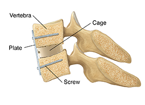
After removing a disk from the front, your surgeon may fuse the vertebrae above and below it. This limits their movement, helping to ease pressure and pain. First, the surgeon cleans the space between the vertebrae, and restores the disk space to its original height before the disk wore out. The surgeon then plugs up the space with a plastic or metal cage. It's filled with bone chips, stem cells, or wedge-shaped bone grafts. A metal plate with screws may be added to the front of the spine to provide stability and increase the rate of fusion. As you heal, the cage or bone graft and the vertebrae above and below will grow together (fuse). After fusion, your ability to bend your neck may be slightly restricted.
 |
| Side view. |
Through the back: posterior approach
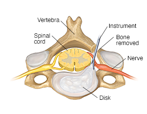
Your surgeon will make an incision (about 2 to 4inches long) in the middle or next to the middle of the back of your neck. Then they may remove bone to reach the problem area. The surgeon then removes the damaged portion of the disk.
 |
| Top view. |
Removing bone
To reach the disk from the back, your surgeon may enlarge the foramina or remove a portion of the lamina. To help ease pressure on the nerves or spinal cord, bone spurs may also be removed. The location and amount of bone removed depend on the type of problem you have.
Through the front: anterior approach
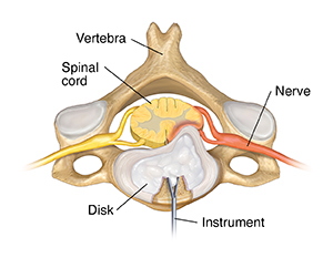
Your surgeon will make a horizontal or vertical incision (about 1 to 3 inches long) on either side of your neck along the neck crease. To reach the disk, soft tissue is moved aside. All or part of the disk that is irritating the nerve is then removed. Your surgeon may remove bone spurs. The vertebrae may then be prepared for a fusion.
 |
| Top view. |
Online Medical Reviewer:
Heather M Trevino BSN RNC
Online Medical Reviewer:
Marianne Fraser MSN RN
Online Medical Reviewer:
Vinita Wadhawan Researcher
Date Last Reviewed:
4/1/2024
© 2000-2025 The StayWell Company, LLC. All rights reserved. This information is not intended as a substitute for professional medical care. Always follow your healthcare professional's instructions.