A
B
C
D
E
F
G
H
I
J
K
L
M
N
O
P
Q
R
S
T
U
V
W
X
Y
Z
Topic IndexLibrary Index
Click a letter to see a list of conditions beginning with that letter.
Click 'Topic Index' to return to the index for the current topic.
Click 'Library Index' to return to the listing of all topics.
Retinopathy of Prematurity
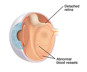
Premature babies are at risk for retinopathy of prematurity (ROP). This is a problem that can affect eyesight. ROP is the growth of abnormal blood vessels on the lining of the back of the eye (retina). In severe cases, the blood vessels can detach the retina from the back of the eye.
What causes ROP?
The blood vessels on the retina don't finish growing until late in pregnancy. When a baby is born prematurely, these blood vessels aren’t yet fully developed. So the blood vessels complete their growth after the baby is born. Factors in the environment outside of the womb may cause them to grow abnormally. One problem may be changing levels of oxygen in the blood. ROP is more likely in younger or smaller preemies.
 |
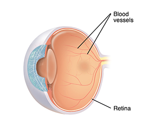
| Normal blood vessels form a delicate web on the retina. |
 |
| With ROP, blood vessels can become large and twisted. They can form a ridge of scar tissue, or pull on the retina, causing it to detach. |
How is ROP diagnosed and monitored?
All premature babies in the NICU (neonatal intensive care unit) have their blood oxygen levels closely monitored. Babies born at or less than 30 weeks and 6 days gestation or weighing 1,500 grams or less (52.5 ounces or less) are examined by an eye care provider (ophthalmologist). Eye exams can also be done by a trained member of the healthcare team using a special camera to look at the back of the eye.
Some babies who weigh between 1,500 and 2,000 grams (52.5 to 70 ounces) and have other health problems may need to have an eye exam because they are also at higher risk for ROP.
During eye exams, drops are used to expand (dilate) the pupil of the eye. This lets the provider look through the pupil to check the blood vessels on the retina. If the provider sees abnormal blood vessels, their ROP is rated from stage 1 (mild) to stage 5 (severe). The location of the blood vessels is also noted.
The first exam may be done around 4 to 8 weeks after birth. Depending on the results of this exam and the baby’s gestational age, they'll need follow-up exams every 1 to 2 weeks.
How is ROP treated?
Mild ROP (stages 1 and 2) often needs no treatment. Babies with moderate to severe ROP may need treatment. Treatment usually depends on the severity of disease. Treatment options are:
-
Laser surgery (laser therapy or photocoagulation). The provider uses beams of light to burn and scar the sides of the retina. This stops abnormal blood vessels from growing and pulling on the retina.
-
Anti-VEGF-therapy. The provider injects anti-VEGF medicine into the inside of the eye (vitreous), near the retina at the back of the eye. This stops abnormal blood vessels from growing and pulling on the retina. This is a newer therapy that is widely used to treat ROP. But, long-term outcomes are still being determined.
-
Scleral buckle. The provider puts a silicone band around the white of the eye (sclera). This band helps push the eye in so that the retina stays along the wall of the eye. The buckle is removed later as the eye grows. If it isn’t removed, a child can become nearsighted. This means they have trouble seeing things that are far away.
-
Vitrectomy. The provider removes the clear gel in the center of the eye (vitreous) and puts saline (salt) solution in its place. The provider can then take out scar tissue, so that the retina doesn’t pull. Only babies with stages 4 or 5 ROP have this surgery.
-
Cryotherapy (freezing). The provider uses a metal probe to freeze and scar the sides of the retina. This prevents spread of abnormal blood vessels and pulling on the retina. This treatment is rarely used now because other therapies generally work better.
Talk with your baby's healthcare provider about which treatment is right for your baby.
What are the long-term effects?
Many babies with ROP have no lasting effects. The more severe the disease, the higher the chance of permanent vision problems. Long-term visual problems occur in 7 in 100 to 3 in 20 children with moderate to severe ROP. ROP can lead to blindness in rare cases.
Most babies will need follow-up eye exams. Babies with ROP are at increased risk for other eye disorders. These include myopia, strabismus, and astigmatism. Your baby may need glasses or other treatments.
Online Medical Reviewer:
Donna Freeborn PhD CNM FNP
Online Medical Reviewer:
Heather M Trevino BSN RNC
Online Medical Reviewer:
Liora C Adler MD
Date Last Reviewed:
3/1/2022
© 2000-2025 The StayWell Company, LLC. All rights reserved. This information is not intended as a substitute for professional medical care. Always follow your healthcare professional's instructions.